Home
/ Back Of Neck Anatomy : Best Head And Neck Anatomy Stock Photos, Pictures ... - Posterior border of the ligament is free, anterior border is attached to the cervical spines and its superior border.
Back Of Neck Anatomy : Best Head And Neck Anatomy Stock Photos, Pictures ... - Posterior border of the ligament is free, anterior border is attached to the cervical spines and its superior border.
Back Of Neck Anatomy : Best Head And Neck Anatomy Stock Photos, Pictures ... - Posterior border of the ligament is free, anterior border is attached to the cervical spines and its superior border.. Muscles of the posterior neck and the back. Beneath the integument the back of neck presents in the median plane the ligamentum nuchae, which is a triangular fibrous sheet and represents upward continuation of supraspinous ligament. The structure is, of course, an important part of the conversation. Whether it's to pass that big test, qualify for that big promotion or even master that cooking technique; This atlas is a comprehensive and affordable learning tool for residents and medical students and especially for radiologists and surgeons.
Anatomical principles underlying cranial nerve lesions; In radiology, the 'head and neck' refers to all the anatomical structures in this region excluding the central nervous system, that is, the brain and spinal co. D) demonstrate sound knowledge of the surface/living and radiological anatomy. The splenius muscles originate at the midline and run laterally and superiorly to their insertions. Despite being a relatively small region, it contains a range of important anatomical features.
Neck anatomy - thyroid | THANC Guide from 2pybk2la9r-flywheel.netdna-ssl.com The neck or cervical spine is the top part of the spine between the head and shoulders. E) demonstrate practical lab skills in anatomy and an appreciation of the ethics. Clinically, surface anatomy is used to split the neck into anterior and posterior triangles which provide clues as to the location of specific structures. The cervical spine has seven vertebra of which the bottom five are designed similarly and the top 2 are very different. Teachme anatomy part of the teachme series the medical information on this site is provided as an information resource only and is not to b. How to view the anatomical labels. The splenius muscles originate at the midline and run laterally and superiorly to their insertions. Despite being a relatively small region, it contains a range of important anatomical features.
Muscles of the posterior neck and the back.
Extends and laterally flexes neck inn: How to view the anatomical labels. D) demonstrate sound knowledge of the surface/living and radiological anatomy. Neck, in land vertebrates, the portion of the body joining the head to the shoulders and chest. The anterior muscles of the neck facilitate swallowing and speech. Dummies has always stood for taking on complex concepts and making them easy to understand. Anatomical principles underlying cranial nerve lesions; Despite being a relatively small region, it contains a range of important anatomical features. The neck is a complex anatomic region between the head and the body. Click now to study the muscles, glands and organs of the neck at kenhub! Neck muscles help support the cervical spine and contribute to movements of the head, neck, upper back, and shoulders. The cervical spine supports the weight and movement of your head and pro. From the sides and the back of the neck, the splenius capitis inserts onto the head region, and the splenius cervicis extends onto the cervical region.
Want to learn more about it? Structures and muscles of the back and neck learn with flashcards, games and more — for free. The neck is the area between the skull base and the clavicles. We've largely focused on the physical aspect of our spinal anatomy in this series. Dummies has always stood for taking on complex concepts and making them easy to understand.
Nerves and arteries of head and neck: Anatomy, branches ... from thumbor.kenhub.com This atlas is a comprehensive and affordable learning tool for residents and medical students and especially for radiologists and surgeons. Despite being a relatively small region, it contains a range of important anatomical features. Click now to study the muscles, glands and organs of the neck at kenhub! How to view the anatomical labels. The neck is the area between the skull base and the clavicles. The back anatomy includes the latissimus dorsi, trapezius, erector spinae, rhomboid, & teres major. An overview of the anatomy of the hand, including the bones of the hand, muscles, blood supply and nerve supply. Anatomy of the head and neck:
Beneath the integument the back of neck presents in the median plane the ligamentum nuchae, which is a triangular fibrous sheet and represents upward continuation of supraspinous ligament.
In order to fully understand primary neck cancers, it helps to understand the anatomy and function of the structures in the neck. Head and neck anatomy is important when considering pathology affecting the same area. Anatomical principles underlying cranial nerve lesions; When to have lower back surgery. The neck or cervical spine is the top part of the spine between the head and shoulders. Muscle head anatomy vocal organ diagram female neck anatomy neck wireframe head neck human anatomy head artery anatomy face pharynx vector neck degree head anatomy 3d. 3d video tutorials and interactive modules on the anatomy of the back including anatomy of the musculature, vertebral column, joints and ligaments. The splenius muscles originate at the midline and run laterally and superiorly to their insertions. Neck muscles help support the cervical spine and contribute to movements of the head, neck, upper back, and shoulders. In radiology, the 'head and neck' refers to all the anatomical structures in this region excluding the central nervous system, that is, the brain and spinal co. The spine runs from the base of your skull down the length of your back, going all the way down to your pelvis. Posterior border of the ligament is free, anterior border is attached to the cervical spines and its superior border. The anterior muscles of the neck facilitate swallowing and speech.
Anatomical principles underlying cranial nerve lesions; Learn about these muscles, their locations & functional the traps are quite a complex set of muscles. An overview of the anatomy of the hand, including the bones of the hand, muscles, blood supply and nerve supply. Clinically, surface anatomy is used to split the neck into anterior and posterior triangles which provide clues as to the location of specific structures. The splenius muscles originate at the midline and run laterally and superiorly to their insertions.
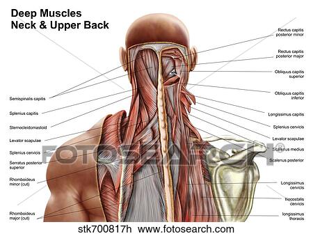
Clip Art of Human anatomy showing deep muscles in the neck ... from fscomps.fotosearch.com Superior angle of scapula a: The neck is the area between the skull base and the clavicles. Learn about these muscles, their locations & functional the traps are quite a complex set of muscles. In order to fully understand primary neck cancers, it helps to understand the anatomy and function of the structures in the neck. The cervical spine has seven vertebra of which the bottom five are designed similarly and the top 2 are very different. Neck muscles help support the cervical spine and contribute to movements of the head, neck, upper back, and shoulders. 3d video tutorials and interactive modules on the anatomy of the back including anatomy of the musculature, vertebral column, joints and ligaments. Dummies has always stood for taking on complex concepts and making them easy to understand.
The anterior muscles of the neck facilitate swallowing and speech.
From the sides and the back of the neck, the splenius capitis inserts onto the head region, and the splenius cervicis extends onto the cervical region. The neck is the area between the skull base and the clavicles. Of working with human remains; Learn everything about the neck anatomy with this topic page. Despite being a relatively small region, it contains a range of important anatomical features. Posterior border of the ligament is free, anterior border is attached to the cervical spines and its superior border. The spine runs from the base of your skull down the length of your back, going all the way down to your pelvis. We've largely focused on the physical aspect of our spinal anatomy in this series. D) demonstrate sound knowledge of the surface/living and radiological anatomy. Your neck is like no other part of the vertebral spinal column and enables your head and neck a wide range of motion. 3d video tutorials and interactive modules on the anatomy of the back including anatomy of the musculature, vertebral column, joints and ligaments. Cervical fascia and interfascial spaces in the neck. Clinically, surface anatomy is used to split the neck into anterior and posterior triangles which provide clues as to the location of specific structures.
